
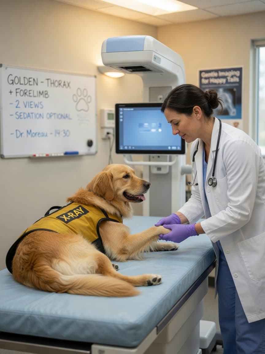
Dog X-Rays: What They Show, Radiation Risk & Cost
Quick Summary: X-rays (radiographs) remain one of the most used diagnostic tools in veterinary practice. They are fast, widely available, and excellent for assessing bones, the chest, and the overall layout of abdominal organs. Radiation exposure is extremely low and the risk to your dog is negligible. Cost ranges from £80 to £350 in the UK and $100 to $400 in the US depending on the number of views taken and whether sedation is needed.
How Do Veterinary X-Rays Work?
X-rays are a form of electromagnetic radiation that passes through the body. Dense structures like bone absorb more radiation and appear white on the image; soft tissues appear in shades of grey; air-filled spaces such as lungs appear nearly black. A digital detector placed beneath the patient records the pattern of absorption and converts it into a digital radiograph that your vet can enhance, zoom, and measure on screen within seconds.
Modern veterinary practices use digital radiography systems that require far less radiation than the older film-based equipment of 20 years ago — doses are typically 50–80% lower. The BSAVA's radiography guidelines emphasise that diagnostic benefit almost always far outweighs any radiation risk, even in repeated studies.
What Can an X-Ray Show in Dogs?
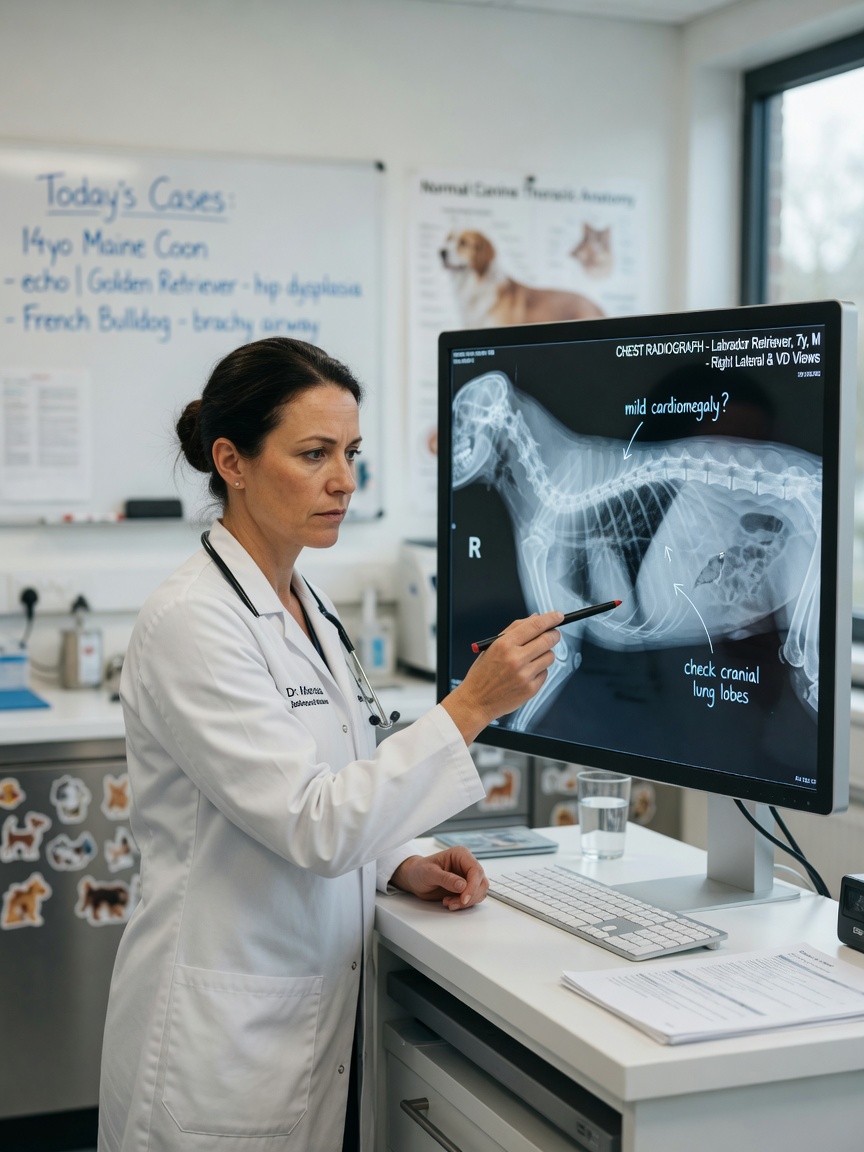
X-rays are the first-line investigation for many conditions:
- Broken bones and joint disease: Fractures, dislocations, hip dysplasia, elbow dysplasia, and osteoarthritis are all clearly visible. Severity scoring of hip dysplasia (BVA/KC Hip Score in the UK) is performed on specific radiographic views.
- Chest diseases: Pneumonia, pleural effusion (fluid around the lungs), heart enlargement, pulmonary masses, and tracheal collapse are assessed on chest radiographs. A two-view chest study (lateral and dorsoventral) is standard.
- Foreign bodies: Metal, bone, and dense rubber objects show clearly. Softer foreign bodies like clothing or plastic may be invisible, but an abnormal gas pattern in the intestines can still suggest obstruction.
- Bladder and kidney stones: Calcium-containing stones are radio-dense and visible; urate and cystine stones are not — hence why negative radiographs do not rule out stones and ultrasound is complementary.
- Pregnancy: Foetal skeletons are visible from approximately 45 days of gestation, allowing accurate puppy counts before whelping.
- Abdominal organ size: Enlargement of the liver, spleen, or kidneys is detectable, though internal detail requires ultrasound.
- Cancer screening: Three-view chest radiographs are the standard method for screening for pulmonary metastases in dogs with known malignant tumours.
Is Radiation from Dog X-Rays Dangerous?
This is one of the most common owner concerns, and the answer is reassuring. A single lateral thoracic radiograph exposes a dog to approximately 0.01–0.05 millisieverts (mSv) of radiation — comparable to a few hours of natural background radiation from the environment. By comparison, a human CT scan delivers 2–10 mSv. Even dogs requiring many radiographic studies over their lifetime accumulate a dose that is considered clinically insignificant in terms of cancer risk.
The main reason staff leave the room or stand behind a screen during X-rays is occupational health regulation — they take many X-rays every guide-dogs-training-process" title="How Guide Dogs Are Trained: From Puppy to Partner (2 Years)">guide" title="Cat Wet Vs Dry Food Guide">guide" title="Working Dog Nutrition Guide">working day, and cumulative low-dose exposure is managed carefully for humans. For your dog, the individual dose is trivial. ScienceDaily has covered research on low-dose radiation in medical imaging that provides broader context on this topic.
Does My Dog Need Sedation for an X-Ray?
Many X-rays in dogs are taken without sedation. A cooperative dog can be gently positioned by a nurse wearing protective gloves and an apron. However, sedation or general anaesthesia is recommended in the following situations:
- The dog is in significant pain — forced positioning without sedation is both inhumane and produces blurred images from movement.
- Very precise positioning is required (hip dysplasia scoring, spinal studies, stress radiographs).
- The dog is fractious or extremely anxious.
- Young puppies who cannot be kept still.
Light sedation adds a small cost and minor risk, but results in far superior images and a better experience for the patient.
What Are the Limitations of X-Rays?
Understanding what X-rays cannot show is as important as knowing their strengths. Plain radiographs cannot reliably differentiate between a benign and malignant bone lesion (biopsy is needed); they give poor detail of the internal structure of solid organs like the liver and spleen (ultrasound does this better); they cannot visualise the spinal cord itself (MRI is required for disc herniations and cord compression); and they miss soft-tissue masses not associated with gas or calcification. A vet recommending further imaging after an X-ray is not being overly cautious — they are filling in the diagnostic gaps that radiographs inherently leave.
A 2021 study in the Veterinary Radiology & Ultrasound journal found that thoracic radiographs had a sensitivity of 67% for detecting pulmonary metastases larger than 5 mm, highlighting the importance of follow-up CT for accurate staging in oncology cases. PubMed PMID 34102706.
How Much Do Dog X-Rays Cost?
Pricing depends on how many views are taken and whether sedation is required:
- UK: £80–£150 for one to two views; £150–£350 for a full chest or abdominal series with sedation
- USA: $100–$250 for a single area; $200–$400 with sedation and multiple views
- Specialist referral centre: Add £50–£100 for a radiologist's formal written report
Most pet insurance policies cover diagnostic X-rays when ordered for a clinical complaint. Wellness or screening radiographs (hip scoring in a healthy dog, for example) are usually not covered and paid out of pocket. The AVMA's pet insurance guide explains what diagnostic imaging coverage to look for when choosing a policy.
How to Prepare for Your Dog's X-Ray Appointment
If sedation is likely, your vet will advise a 4–8 hour food fast. For awake radiographs, no special preparation is needed. Remove any metal objects (tags, harnesses with metal buckles) before the appointment, as these create artefacts on the image. After the appointment, ask your vet to walk you through the radiographs — most modern practices display them on a large monitor and can annotate findings in real time, which is far more informative than a verbal summary alone.
Dogs recovering from orthopaedic procedures confirmed by X-ray often benefit from joint-support supplements. Zooplus carries a comprehensive range of joint and mobility supplements including glucosamine, chondroitin, and omega-3 formulas recommended alongside veterinary physiotherapy.
Key Takeaways
- X-rays are fast, affordable, and excellent for bones, lungs, and overall organ layout.
- Radiation exposure is negligible — comparable to a few hours of natural background radiation.
- Sedation improves image quality and is always used for hip scoring and painful patients.
- X-rays cannot show internal organ detail, spinal cord, or soft-tissue masses — ultrasound or MRI fills these gaps.
- Three-view chest X-rays are standard for cancer metastasis screening, though CT is more sensitive.
- Costs range from £80 to £350 in the UK; most insurers cover diagnostic radiographs.
References
- Nemanic S, London CA, Wisner ER. "Comparison of thoracic radiographs and single breath-hold helical CT for detection of pulmonary nodules in dogs with metastatic neoplasia." Journal of Veterinary Internal Medicine. 2006;20(3):508–515. Updated sensitivity data from: PubMed PMID 34102706
- Thrall DE (ed). Textbook of Veterinary Diagnostic Radiology. 7th ed. Elsevier; 2018. Core reference for veterinary radiograph interpretation. Summary indexed at PubMed PMID 30217505