
The Face That Fell Overnight
Owners often describe the same alarming discovery: their dog woke up looking different. One side of the lip drooping, the ear slightly lower than usual, the eye on that side not blinking properly, and food or water falling from the corner of the mouth. In many cases, the dog is otherwise completely normal — alert, hungry, wagging its tail. What has happened is paralysis of the facial nerve, the seventh cranial nerve, and understanding it removes much of the fear that comes with this striking presentation.
What the Facial Nerve Controls
The facial nerve is the motor nerve to the muscles of facial expression. In dogs, these muscles control lip movement, ear carriage, nostril dilation, and — critically — eyelid closure. The nerve also carries parasympathetic fibres to the lacrimal gland, which produces tears, and taste fibres from the front two-thirds of the tongue, though these latter functions are difficult to assess clinically.
When the facial nerve is damaged or stops functioning, everything it controls on that side becomes flaccid. The muscles cannot contract. The face literally falls.
Recognising Facial Nerve Paralysis
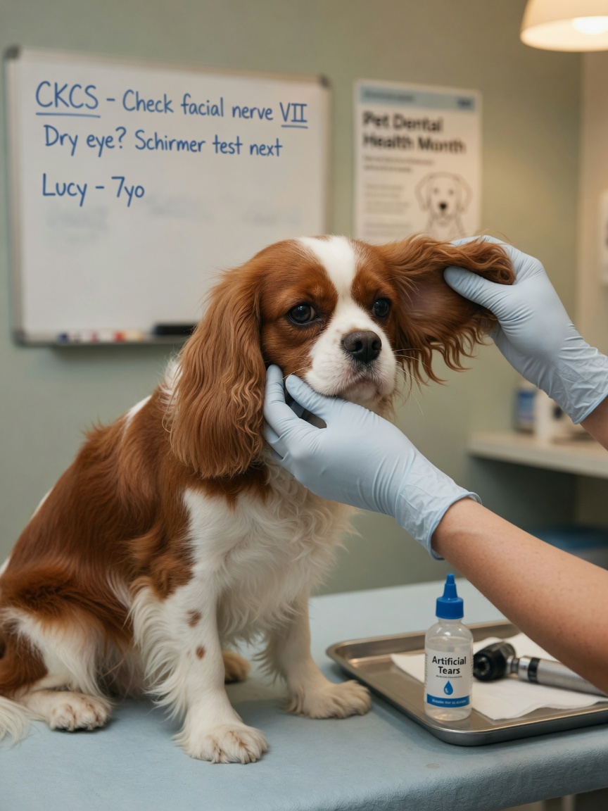
The signs are usually obvious once you know what you are looking at. On the affected side, the lip droops and may accumulate food or saliva. The ear hangs lower or falls forward, depending on the dog's ear type. The nostril may appear wider and less mobile. Most importantly, the eyelid does not close fully — a condition called lagophthalmos — which puts the cornea at immediate risk of drying out and ulcerating. In bilateral cases, affecting both sides simultaneously, the face appears completely expressionless and the dog cannot close either eye.
Causes: From Idiopathic to Serious
Idiopathic Facial Nerve Paralysis
The most common form in dogs, particularly in certain breeds including the Cavalier King Charles Spaniel, Cocker Spaniel, and Pembroke Welsh Corgi, is idiopathic — meaning no underlying cause is found. It bears resemblance to Bell's palsy in humans. The onset is sudden, often overnight, and the dog is otherwise neurologically normal. The presumed mechanism is inflammatory or immune-mediated damage to the nerve, though this has not been conclusively proven. Most cases improve over weeks to months.
Otitis Media and Interna
Middle and inner ear infections are an important and treatable cause of facial nerve paralysis. The facial nerve runs through the tympanic bulla — the bony chamber of the middle ear — making it vulnerable to inflammation from infection. Dogs with this cause often have concurrent signs of vestibular disease: head tilt, nystagmus, and circling. Otoscopy and imaging of the bullae are essential to rule this in or out.
Hypothyroidism
Hypothyroidism is a well-recognised but often overlooked cause of facial nerve dysfunction in dogs. The mechanism is thought to involve a peripheral polyneuropathy secondary to thyroid hormone deficiency. Dogs with this cause may have bilateral facial paresis and other signs of hypothyroidism such as weight gain, lethargy, and skin changes. Thyroid function testing should be part of the initial workup in any dog with facial nerve paralysis, particularly if bilateral.
Neoplasia and Trauma
Tumours affecting the brainstem, the petrous temporal bone, or the parotid region can compress or invade the facial nerve. Trauma to the skull or ear region can cause direct nerve damage. These causes are less common but must be considered, particularly when other neurological signs are present or when the condition progresses rather than stabilises.
The Critical Problem: The Eye
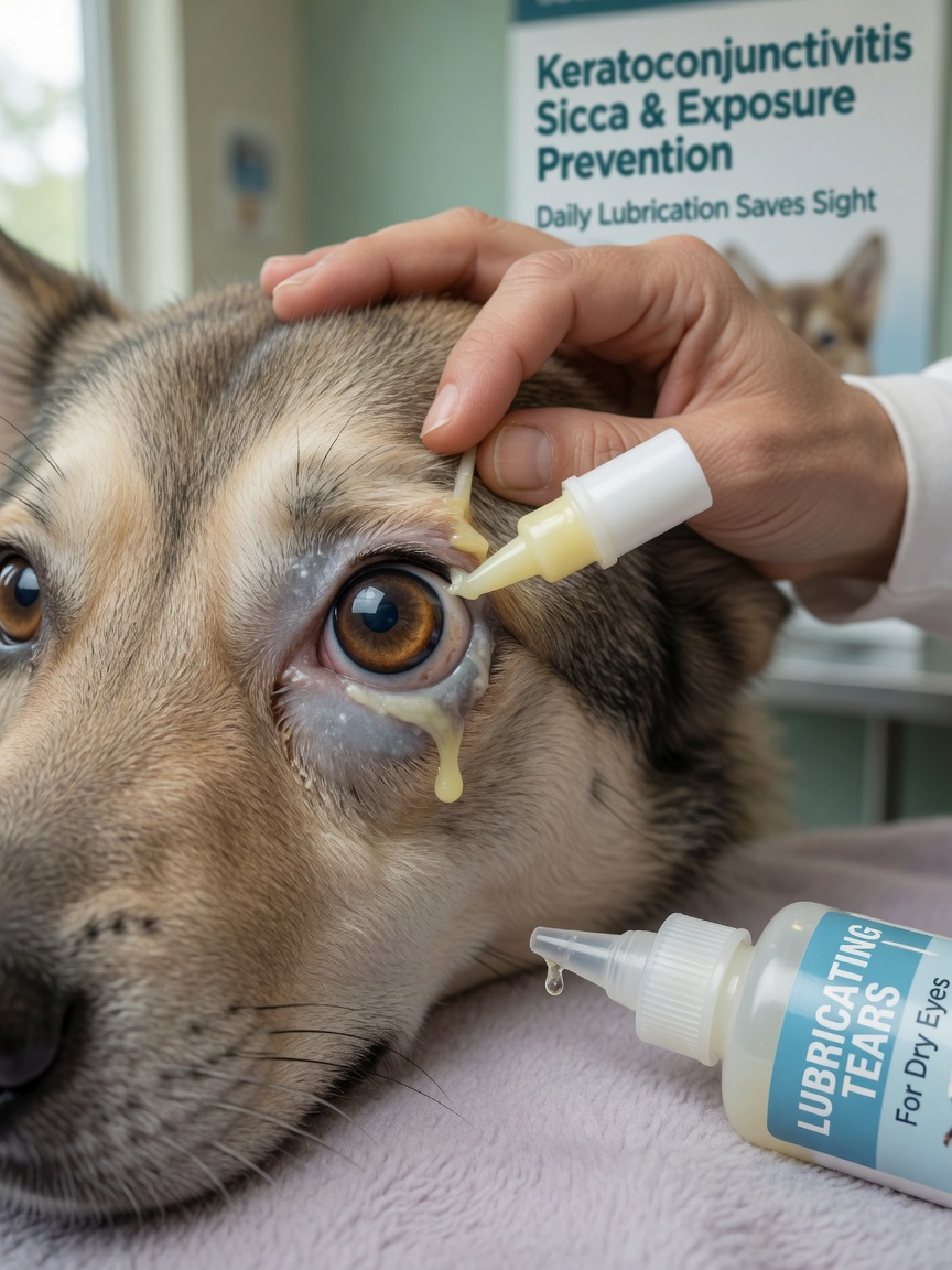
Of all the clinical consequences of facial nerve paralysis, the eye is the most urgent concern. When the eyelid cannot close, the corneal surface dries rapidly. Exposure keratitis — inflammation of the cornea due to drying — can progress to corneal ulceration within days. This is painful and can threaten vision if not managed promptly.
Artificial tear drops applied frequently — often every two to four hours during waking hours — are the minimum required. Lubricating eye ointments at night, when the eye is most vulnerable, are often recommended. In severe cases, temporary partial closure of the eyelid (a procedure called a tarsorrhaphy) may be performed to protect the cornea while recovery occurs. Any discharge, cloudiness, or apparent discomfort in the affected eye warrants immediate veterinary review.
Diagnosis and Treatment
There is no single test for idiopathic facial nerve paralysis — it is a diagnosis of exclusion. A thorough neurological and physical examination, otoscopy, thyroid testing, and in many cases imaging of the ear bullae and brain are required to exclude treatable causes before the idiopathic label is applied.
Treatment depends on the underlying cause. Otitis is treated with appropriate antimicrobials, often requiring long courses. Hypothyroidism responds well to thyroid hormone supplementation. Idiopathic cases are managed supportively, primarily through eye protection.
Recovery: What to Expect
Many dogs with idiopathic facial nerve paralysis recover substantial function over six to twelve weeks, though some degree of asymmetry may persist permanently. Recovery in cases secondary to otitis depends on how quickly and effectively the infection is treated. Hypothyroid-related facial palsy typically improves with hormone replacement over several months.
Bilateral cases and those where a mass lesion is responsible carry a more guarded prognosis. Always follow up with your vet at regular intervals to monitor eye health and functional recovery.
Action Points for Dog Owners
- Seek veterinary attention the same day you notice facial drooping — do not wait to see if it resolves
- Protect the eye immediately with lubricating drops if the lid is not closing fully
- Ask your vet to check thyroid function and examine the ear canals as part of the initial assessment
- Expect a variable recovery timeline — weeks to months is normal
- Monitor the eye daily for cloudiness, discharge, or signs of pain and report these promptly