
What Are Mast Cell Tumours?
Mast cell tumours are the most common malignant skin tumour in dogs, accounting for approximately 20 percent of all skin masses diagnosed. Mast cells are a normal component of the immune system, found in connective tissue throughout the body and particularly concentrated in the skin, respiratory tract, and digestive system. They play a role in allergic responses and wound healing by releasing chemicals such as histamine and heparin. When mast cells undergo malignant transformation and proliferate abnormally, a mast cell tumour — also referred to as a mastocytoma — forms.
These tumours can occur in dogs of any age or breed, but certain breeds carry a significantly higher predisposition. Boxers, Bulldogs, Pugs, Boston Terriers, Labrador Retrievers, Golden Retrievers, and Weimaraners are among those most commonly affected. Boxers in particular are notorious for developing mast cell tumours, though they tend to have a more favourable grade and prognosis than those seen in some other breeds.
What Do They Look Like?
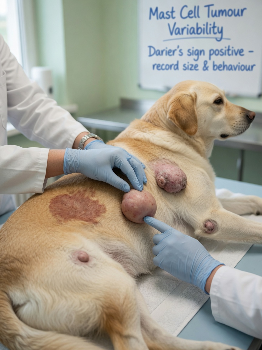
This is where mast cell tumours earn their reputation as the great imitators. They can look like almost anything — a small raised lump, a flat skin discolouration, a hairless patch, or a large, angry, ulcerated mass. They may be firm or soft, slow-growing or rapidly expanding. Some fluctuate in size, appearing to shrink one week and enlarge the next, a phenomenon caused by degranulation of the mast cells releasing histamine.
Darier's sign — reddening and swelling of the mass when handled — is a useful clinical clue, as mechanical stimulation of mast cells causes them to degranulate locally. However, this sign is not reliably present and its absence does not rule the diagnosis out.
Because of their deceptive appearance, the veterinary advice is consistent: any new lump on a dog should be investigated promptly rather than assumed to be benign based on how it looks. The only reliable way to know what a lump is involves sampling it.
Diagnosis: Fine Needle Aspirate and Beyond
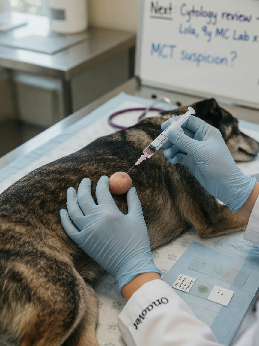
The first step in investigating a suspicious mass is typically a fine needle aspirate (FNA), a quick, minimally invasive procedure performed in the consulting room. A needle is inserted into the mass and cells are collected onto a slide for cytological examination. Mast cell tumours are usually readily identifiable on cytology due to the characteristic granules within the cells.
Once a mast cell tumour is confirmed, surgical removal with histopathology is the standard next step. The excised tissue is submitted to a pathologist who assigns a grade — traditionally Patnaik grades I, II, or III, or using the more recent Kiupel two-tier system (low-grade and high-grade). Grade is the single most important prognostic indicator, influencing treatment decisions and expected outcomes significantly.
Additional staging investigations may include lymph node aspirates, abdominal ultrasound, chest radiographs, and in some cases a buffy coat smear to look for circulating mast cells. The extent of staging required depends on the grade of the tumour and clinical findings at the time of examination.
Treatment Approaches
Surgery with wide, clean margins remains the primary treatment for localised mast cell tumours. The goal is to remove the tumour with a margin of normal tissue on all sides, reducing the risk of local recurrence. When wide margins are not achievable due to location — on the face, feet, or near joints, for example — radiation therapy may be recommended as an adjunct.
Higher-grade tumours, those with incomplete excision, or cases with evidence of spread may be managed with chemotherapy. Vinblastine and lomustine are commonly used agents. Tyrosine kinase inhibitors such as toceranib (Palladia) and masitinib (Masivet) are targeted oral therapies licensed for use in dogs and have demonstrated efficacy particularly in tumours harbouring KIT mutations.
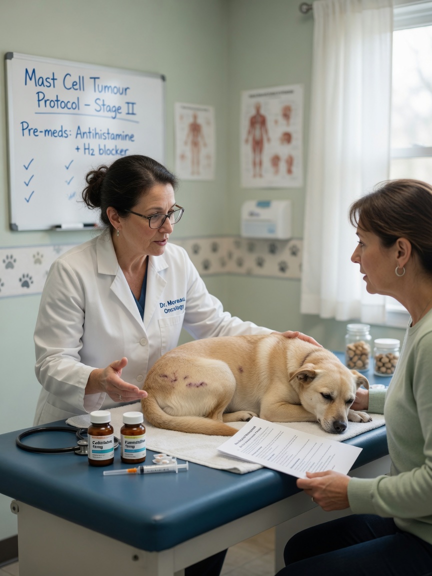
Because mast cell degranulation releases histamine, pre- and post-surgical management often includes antihistamines (both H1 and H2 blockers such as diphenhydramine and famotidine) and sometimes a short course of prednisolone, to reduce the risk of gastrointestinal ulceration from histamine release during handling of the tumour.
Prognosis and Follow-Up
The prognosis for mast cell tumours is highly variable and depends on grade, stage, location, and completeness of surgical excision. Low-grade tumours completely excised surgically carry an excellent prognosis, with many dogs considered cured following surgery alone. High-grade tumours carry a significantly more guarded outlook, with median survival times measured in months even with treatment.
Regular follow-up is essential. Veterinary oncologists typically recommend recheck examinations every one to three months for the first two years following treatment, as mast cell tumours can recur locally or develop at new sites. Early detection of any new masses gives the best chance of successful management.
It is understandably anxiety-inducing to discover a lump on your dog, but early investigation genuinely makes a difference. Many dogs with mast cell tumours go on to live comfortably for years with appropriate management.