
A Parasite That Can Turn Into a Tumour
Spirocerca lupi is not a household name among dog owners in Northern Europe — but for pets that have lived in or travelled through Southern Europe, the Middle East, Africa, Asia, or the Americas, it represents a serious and often overlooked risk. This parasitic nematode takes up residence in the oesophageal wall of dogs, causing granulomas that, in a significant proportion of cases, undergo malignant transformation into sarcomas. It is one of the very few known examples of a parasite directly inducing cancer in a companion animal.
Life Cycle: A Complex Journey Through Multiple Hosts
The life cycle of S. lupi involves obligate intermediate hosts and is considerably more elaborate than that of many common canine parasites.
The Dung Beetle as Intermediate Host
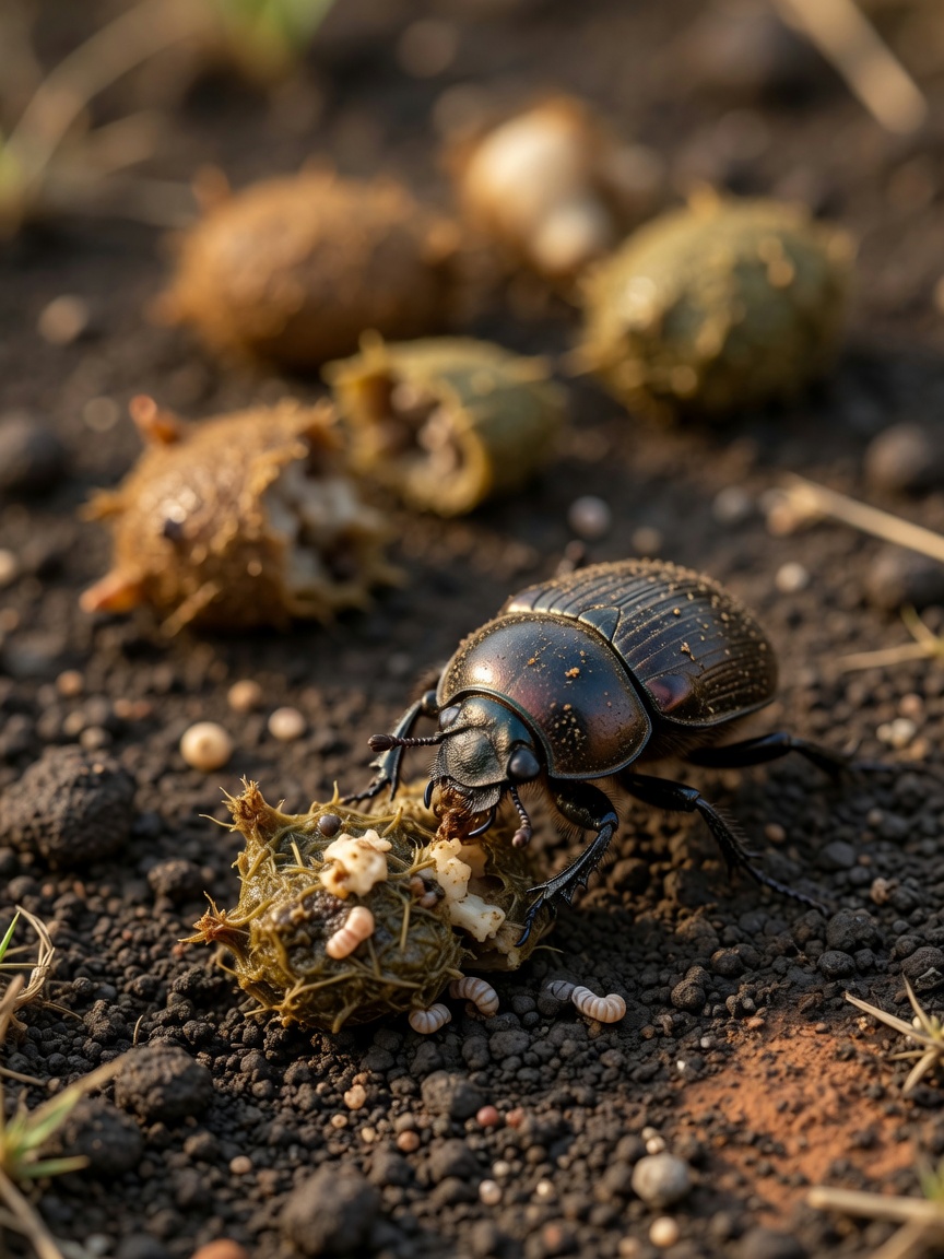
Adult worms living in oesophageal nodules shed eggs into the faeces of the definitive host. Coprophagous beetles — dung beetles — ingest these eggs, and larvae develop to the infective third stage within the beetle. Dogs become infected by ingesting an infected beetle directly, or by eating a paratenic host: a reptile, bird, small mammal, or hedgehog that has itself consumed an infected beetle. This indirect transmission route makes control particularly challenging.
Migration Inside the Dog
Following ingestion, L3 larvae penetrate the gastric wall and migrate along the wall of the aorta — sometimes causing aortic aneurysms or scarring in the process — before eventually reaching the caudal oesophagus. Here, they encyst in the submucosa and muscularis, forming characteristic nodules. The prepatent period is approximately six months.
Clinical Signs and Why They Are So Varied
Spirocercosis can present in several distinct ways depending on which structures are affected and whether malignant transformation has occurred.
Oesophageal Signs
The most common presentation involves signs relating to the oesophageal nodules: regurgitation, vomiting, hypersalivation, and difficulty swallowing. Dogs may lose weight despite a maintained appetite as food fails to pass normally. Oesophageal obstruction or perforation can occur in severe cases.
Respiratory and Vascular Complications
Migration along the aorta can cause aortic scarring and, rarely, life-threatening rupture. Larvae occasionally migrate to the spinal cord, lungs, or trachea, producing neurological signs or respiratory distress. Pulmonary osteopathy — periosteal new bone formation along the limbs — is a paraneoplastic sign that appears when sarcomatous transformation has occurred and is a useful clinical clue.
Spondylitis
Ventral spondylitis of the caudal thoracic vertebrae, visible on radiographs, is a well-recognised feature of spirocercosis and should prompt further investigation in dogs with appropriate travel history.
Diagnosis
No single diagnostic test is pathognomonic. The index of suspicion must be high in dogs from endemic regions presenting with oesophageal signs or unexplained weight loss.
- Faecal flotation using a specific high-density solution may identify the characteristic L-shaped larvated eggs, but sensitivity is low
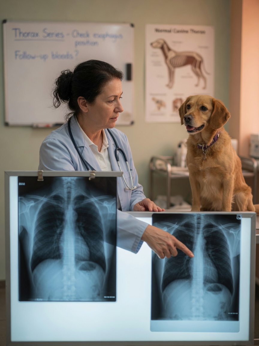
- Thoracic radiography may reveal oesophageal masses, aortic irregularities, vertebral changes, or hypertrophic osteopathy
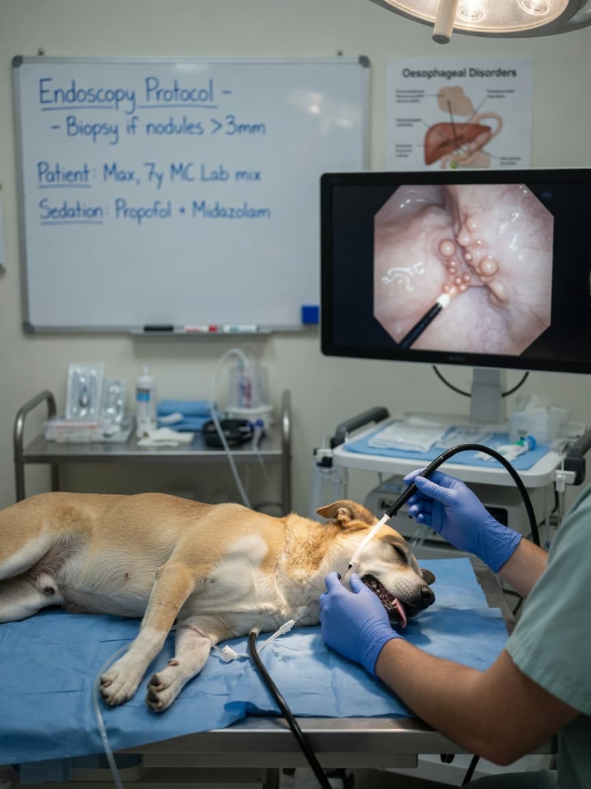
- Oesophagoscopy is the most sensitive diagnostic tool, allowing direct visualisation and biopsy of nodules
- CT imaging provides detailed assessment of lesion extent and vascular involvement
Biopsy of oesophageal nodules is essential to distinguish benign granulomas from sarcomatous transformation, as this distinction is critical for prognosis and treatment planning.
Treatment Options
The macrocyclic lactone doramectin, administered as repeated subcutaneous injections, is the most widely studied treatment protocol for spirocercosis and has demonstrated efficacy in eliminating worms and reducing nodule size. Milbemycin oxime has also been used with some success.
Where malignant transformation has occurred, the prognosis is considerably more guarded. Surgical resection of sarcomatous oesophageal masses is technically demanding and associated with high complication rates. Adjunctive chemotherapy protocols have been explored at specialist centres, but outcomes for malignant spirocercosis remain poor.
Treatment should always be supervised by a veterinarian experienced in the disease, ideally with access to endoscopic monitoring to assess nodule response over time.
Prevention and the Travelling Dog
Preventing access to intermediate and paratenic hosts is the cornerstone of prevention but is difficult to achieve completely in practice. Dogs that hunt, scavenge, or roam freely in endemic areas face the greatest risk. Some veterinarians in endemic regions use prophylactic doramectin administration, though evidence-based protocols for primary prevention in travelling dogs are not yet standardised.
Owners planning to travel with dogs to Southern Africa, the Mediterranean basin, the Middle East, or South and Southeast Asia should discuss S. lupi risk with their veterinarian well in advance. Dogs returning from endemic regions with any history of regurgitation, unexplained weight loss, or vomiting should have spirocercosis included in the differential diagnosis.
A Summary for Owners of Travelling Dogs
- Spirocerca lupi is endemic across much of Africa, Asia, Southern Europe, and parts of the Americas
- Infection occurs through ingestion of beetles or paratenic hosts — not from other dogs
- Oesophageal nodules can undergo malignant transformation into sarcoma
- Clinical signs include regurgitation, weight loss, and hypertrophic osteopathy
- Endoscopy is the most sensitive diagnostic tool; radiography provides useful supporting evidence
- Treatment with macrocyclic lactones is effective for benign disease but requires veterinary supervision and monitoring
- Discuss prevention with your veterinarian before any travel to endemic regions
Spirocercosis is preventable, treatable in its early stages, and potentially fatal if missed. For dogs with a history of international travel, keeping this parasite on the diagnostic radar is not optional — it is responsible ownership.