
When the Spring in the Step Breaks
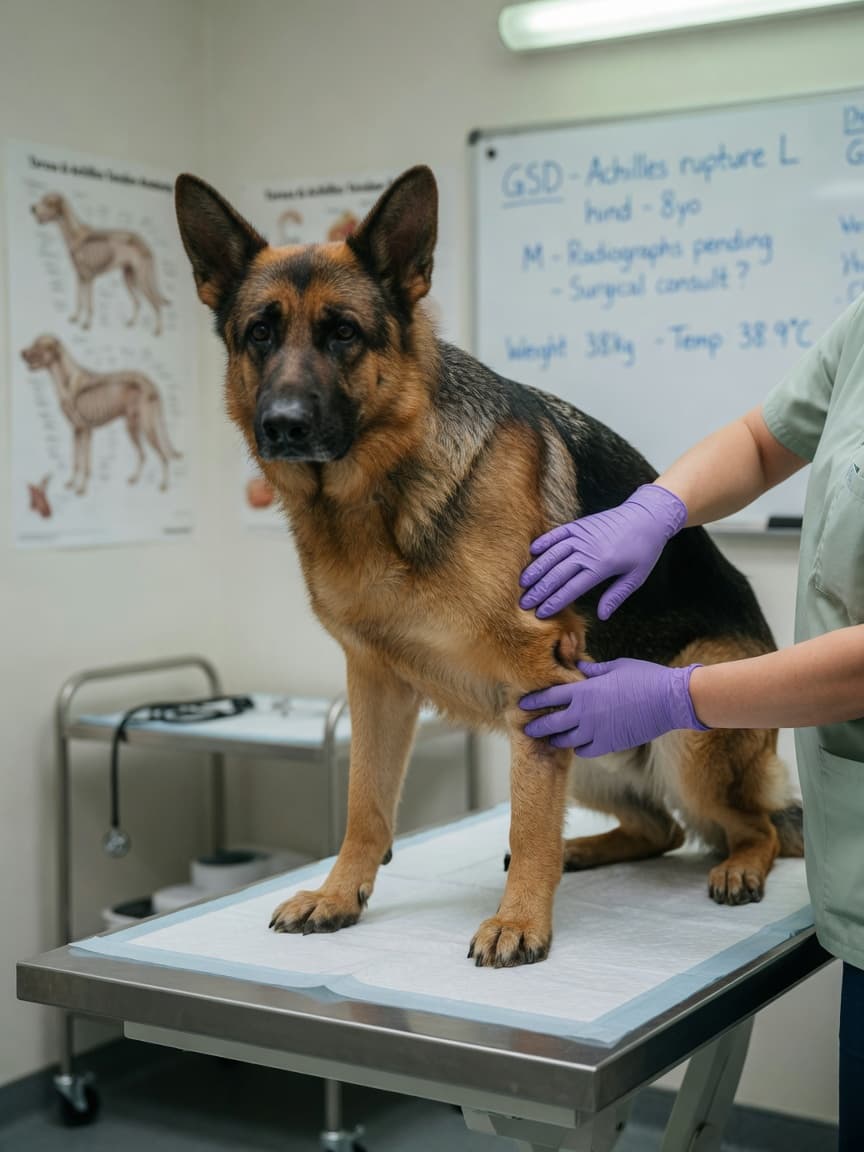
Watch a dog moving at speed and you will notice the characteristic springy, powerful push-off from the hindlimbs. That elastic energy is largely generated by the common calcaneal tendon — commonly called the Achilles tendon — the thick fibrous band connecting the calf muscles to the heel bone (calcaneus). When this structure is partially or completely ruptured, the result is immediately apparent: a dog that walks with a dropped heel, an over-flexed hock, and a distinctive plantigrade stance, appearing almost to walk flat-footed on the lower limb. Without prompt treatment, the consequences for long-term function are serious.
The Anatomy of the Canine Achilles Tendon
In dogs, the common calcaneal tendon is not a single structure but a combination of five tendons, the largest of which is the gastrocnemius tendon. The superficial digital flexor tendon, and tendons from the gracilis, biceps femoris, and semitendinosus muscles also contribute. This complex inserts onto the tuber calcanei — the point of the heel — and is responsible for extending the hock during weight-bearing and propulsion.
Injury can affect one or more components of this tendon complex. A complete rupture of the gastrocnemius component produces the classic dropped hock and plantigrade stance. Partial tears can be more subtle and are sometimes missed on initial examination.
Causes of Achilles Tendon Rupture
Acute Traumatic Rupture
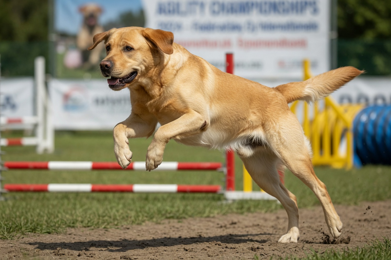
Sudden, high-force incidents — a misjudged jump, an awkward landing, or a penetrating wound — can rupture the tendon acutely. In these cases the onset of lameness is immediate, and the clinical signs are often dramatic. Racing and agility dogs are at elevated risk due to the extreme forces placed on the Achilles tendon during high-speed activity.
Degenerative Tendinopathy
In middle-aged to older dogs, the Achilles tendon can weaken progressively through degenerative change — a process known as tendinopathy. The internal structure of the tendon becomes disorganised, blood supply diminishes, and the tendon's tensile strength declines. Rupture can then occur with what appears to be minimal force: a normal walk or a routine movement. Labrador Retrievers and Dobermanns are among the breeds reported more frequently with degenerative Achilles tendon disease, though any dog can be affected.
Steroid-Associated Weakening
Repeated peritendinous corticosteroid injections — sometimes used to manage local inflammation — are associated with tendon weakening and can predispose to rupture. This is a recognised risk that should inform clinical decision-making around tendon management.
Diagnosis
A veterinarian can usually diagnose Achilles tendon rupture through physical examination alone. The plantigrade stance, dropped hock, and abnormal flexibility of the hock on palpation are strongly indicative. Tensioning the tendon manually — by extending the stifle — should produce hock extension in an intact tendon; absence of this response confirms significant disruption.
Diagnostic imaging confirms the extent of injury. Ultrasound is the modality of choice for tendon assessment, allowing direct visualisation of the tendon fibres, the location and extent of disruption, and any associated fluid or swelling. MRI provides more detailed soft tissue information and may be used in complex or ambiguous cases. Radiographs are taken to exclude concurrent bony injury, including avulsion fractures at the calcaneal insertion.
Surgical Repair
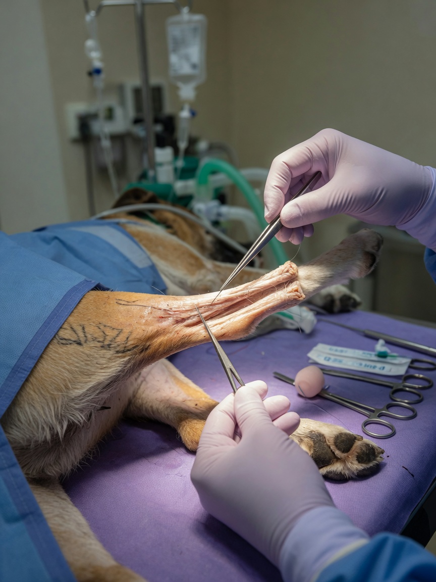
Complete Achilles tendon rupture requires surgical repair. Non-surgical management is occasionally considered for partial tears in specific circumstances, but most cases — and all complete ruptures — necessitate surgical intervention to restore tensile continuity and achieve functional recovery.
Surgical Techniques
The surgeon identifies the torn tendon ends, debrides any devitalised tissue, and appositions the ends using suture techniques designed to withstand the substantial forces the Achilles tendon must bear. Common repair patterns include the three-loop pulley, the Bunnell, and the locking-loop suture configurations, chosen for their mechanical strength in tendon tissue. In cases of significant tissue loss, augmentation with synthetic implants or autologous tissue grafts may be required.
Following tendon repair, the hock is immobilised in extension using an external coaptation device — typically a splint or a hobble bandage system — to protect the repair while initial healing occurs. The period of immobilisation usually spans six to eight weeks.
Complications
Tendon repairs are technically demanding, and complications are not uncommon. Re-rupture, infection, implant failure, and wound breakdown are all recognised risks. Skin and wound complications are of particular concern over the hock, where skin is under tension and blood supply is less abundant. Close post-operative monitoring and bandage management are essential.
Rehabilitation and Long-Term Outlook
Recovery from Achilles tendon repair is a prolonged process. Tendons heal slowly — the remodelling phase of tendon repair continues for months after the initial healing. Exercise restriction during the first two months is strict, after which controlled physiotherapy begins. Hydrotherapy with underwater treadmill work is particularly valuable for rebuilding hindlimb muscle without placing the tendon under excessive load.
Controlled lead walks are gradually extended from around eight weeks, and return to full activity typically occurs at four to six months in uncomplicated cases. The owner's commitment to the rehabilitation programme is a major determinant of outcome. Dogs that recover well often return to comfortable, functional activity, though return to competitive sport is variable and depends on the degree of initial injury and the quality of repair.
Key Points for Owners
- Achilles tendon rupture presents as a dropped hock and plantigrade stance — seek veterinary assessment immediately if you notice this.
- Both traumatic and degenerative causes exist; middle-aged large-breed dogs are at particular risk of degenerative rupture.
- Surgical repair is required for complete ruptures and most significant partial tears.
- Post-operative immobilisation and dedicated physiotherapy are essential components of recovery.
- Recovery to functional activity takes four to six months in uncomplicated cases.
- Early veterinary assessment improves the likelihood of a successful outcome — delayed treatment allows tendon ends to retract and increases surgical complexity.