
One Morning Everything Changes
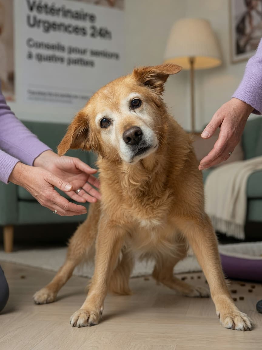
You wake up to find your dog unable to stand, head tilted sharply to one side, eyes flickering rapidly back and forth. Your first thought is stroke or brain tumour. You rush to the vet, braced for devastating news. The diagnosis comes back: idiopathic vestibular disease. Most dogs recover almost completely within two to three weeks. This is the condition that terrifies owners and then, in the majority of cases, largely resolves on its own — but understanding it properly matters, because not every presentation is benign.
What Is the Vestibular System?
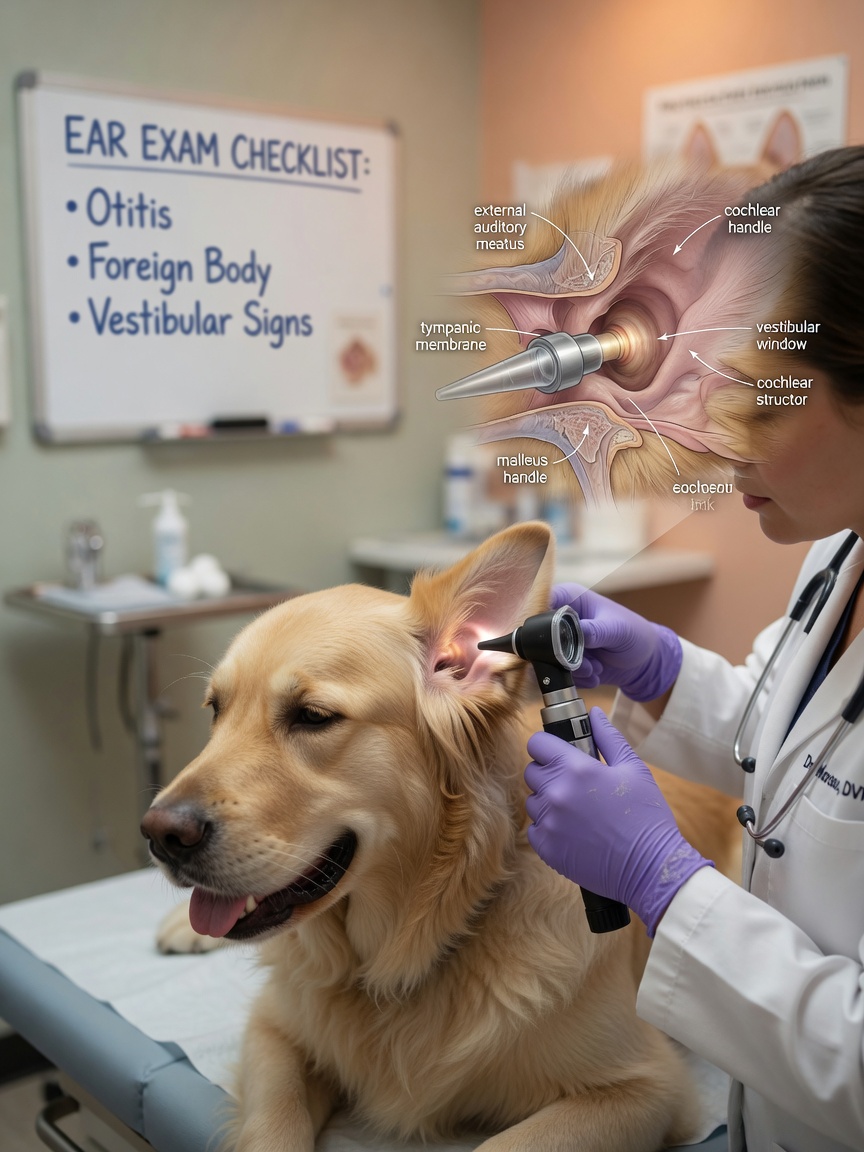
The vestibular system is the body's balance and spatial orientation network. It consists of sensory structures in the inner ear and a processing centre in the brainstem, connected by the vestibular nerve. This system continuously tells the brain where the head is in space and coordinates the muscular responses that keep the body upright. When it malfunctions, the result is dramatic: the dog experiences a sensation similar to severe vertigo, and the body's compensatory systems go haywire.
The Two Types: Peripheral and Central
The clinical distinction between peripheral and central vestibular disease is critical, because they carry very different prognoses.
Peripheral Vestibular Disease
Peripheral disease originates in the inner ear or vestibular nerve. It is the more common form and, crucially, the more benign. Idiopathic vestibular disease — where no underlying cause can be found — falls into this category and is particularly prevalent in older dogs, earning the colloquial name "old dog vestibular syndrome." Other peripheral causes include inner ear infections (otitis interna), middle ear disease, polyps, or adverse reactions to certain medications, particularly some aminoglycoside antibiotics.
Central Vestibular Disease
Central disease involves the brainstem itself and carries a more serious prognosis. Causes include brain tumours, inflammatory brain disease (encephalitis), strokes, and thiamine deficiency. Distinguishing central from peripheral disease is therefore essential and cannot always be done on clinical signs alone — imaging is frequently required.
Signs to Know
The hallmark signs of vestibular disease, regardless of cause, include:
- Sudden onset of loss of balance and falling, often to one side.
- Head tilt — the head rotates so one ear drops lower than the other, typically towards the affected side.
- Nystagmus — rapid, involuntary flickering of the eyes, usually from side to side (horizontal) or in a rotary pattern.
- Ataxia — a staggering, uncoordinated gait as though the dog is profoundly drunk.
- Nausea and vomiting, which can be severe in the acute phase due to the vertigo-like sensation.
- Reluctance to move or eat in the first 24 to 48 hours.
Features that raise concern for central rather than peripheral disease include vertical nystagmus (eyes flickering up and down), pronounced weakness or paralysis of limbs, difficulty swallowing, facial paralysis, or marked changes in mentation. Any of these warrant urgent veterinary assessment.
Diagnosis: When Is Imaging Needed?
Many cases of peripheral vestibular disease can be diagnosed clinically, particularly when the onset is sudden, the dog is older, and the neurological examination does not reveal signs pointing to central involvement. Ear examination to exclude otitis media or interna is standard.
When the diagnosis is uncertain, when signs point towards central disease, when there is no improvement after 72 hours, or when the dog deteriorates, MRI is indicated. Blood tests and urine analysis are typically performed to rule out systemic illness. In cases where inner ear infection is suspected without imaging, deep ear swabs and culture guide antibiotic choice.
Treatment and Recovery
Idiopathic Vestibular Disease
There is no specific treatment for the idiopathic form — it resolves on its own. Management is supportive: anti-nausea medication (maropitant is widely used in veterinary practice) helps during the acute vomiting phase. Some dogs benefit from mild sedation in the first day or two if distress is significant. Owners need to ensure the dog is kept safe — padded environments to prevent injury from falling, hand-feeding if the dog cannot reach a bowl, and help with outdoor toileting.
Improvement typically begins within 72 hours. Most dogs show substantial recovery within one to two weeks. A residual head tilt may persist permanently in some dogs but does not impair quality of life. Nystagmus resolves as the brain compensates for the vestibular asymmetry.
Underlying Causes
When a cause is identified, treatment targets it directly. Inner ear infections require systemic antibiotics for a minimum of six to eight weeks. Inflammatory brain disease requires immunosuppressive therapy. Cerebrovascular disease management focuses on identifying and addressing risk factors such as hypertension, hypothyroidism, or hyperadrenocorticism.
Supporting Your Dog Through Recovery
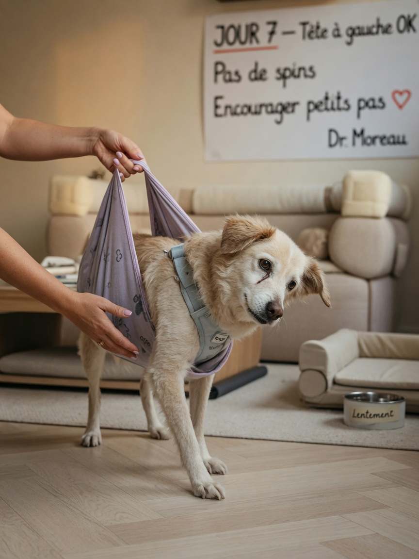
- Create a safe, confined space with padded flooring to prevent injury during the unsteady phase.
- Offer water frequently, using a raised bowl or by hand if the dog struggles to reach down.
- Carry dogs on stairs — do not let them attempt steps unsupervised until balance has substantially returned.
- Provide short, supported walks outdoors for toileting; a towel or commercial support harness can assist larger dogs.
- Avoid sudden position changes and keep the environment calm and quiet.
- Contact your vet if there is no improvement after 72 hours, if signs worsen, or if the dog cannot keep water down.
Vestibular disease can look catastrophic in the moment. For most older dogs, the prognosis is genuinely good — the condition improves without aggressive intervention, and watching the recovery unfold, from a dog that cannot stand to one strolling normally within a fortnight, is one of veterinary neurology's more gratifying outcomes. That said, always have a vet assess the episode promptly to confirm the diagnosis and rule out the less benign causes that require specific treatment.